Revista Científica
versión impresa ISSN 0798-2259
Rev. Cient. (Maracaibo) v.19 n.6 Maracaibo dic. 2009
Accuracy of ultrasonographic measurements of kidney dog for clinical use
Exactitud de las medidas ultrasonográficas renales en el perro para uso clínico
Rafael Barrera, Javier Duque, Patricia Ruiz y Concepción Zaragoza
Department of Medicine and Animal Health, Faculty of Veterinary Sciences, University of Extremadura, Avda. Universidad s/n, 10071 Caceres, Spain. Corresponding author: Concepción Zaragoza. Departamento de Medicina y Sanidad Animal. Facultad de Veterinaria. Universidad de Extremadura. Avda. de la Universidad s/n. 10071- Caceres, Spain. Phone: 34 927257162. Fax number: 34 927257102. E-mail: zaragoza@unex.es - rabacha@unex.es
ABSTRACT
The aim of this study was to determine the accuracy of ultrasonographic measurement of canine kidneys. A method agreement analysis comparing pairs of measurements (ultrasonographic kidney dimensions and direct kidney dimensions) was performed. Nineteen dogs aging from one to thirteen years were used. The ultrasonographic and anatomic linear parameters obtained from the kidneys were the following: width, length and height; the kidney volume was estimated from these measures using the formula of an ellipsoid. The statistical method performed (method agreement) demonstrated a satisfactory agreement. It can be concluded that ultrasound measurement is sufficiently accurate for clinical use, assuming a careful scanning technique.
Key words: Kidney, dog, ultrasonography, measurements, statistical agreement analysis.
RESUMEN
El objetivo de este trabajo fue determinar la fiabilidad de las medidas ecográficas de los riñones del perro. Se llevó a cabo un análisis de concordancia entre métodos (medidas ecográficas del riñón y medidas anatómicas directas). Se utilizaron perros cuyas edades iban de uno a trece años. Las medidas ecográficas y anatómicas lineales que se obtuvieron de los riñones fueron las siguientes: anchura, longitud y altura. El volumen renal se estimó aplicando estas medidas en la fórmula de un elipsoide. A través de un análisis de concordancia estadística se observó un nivel de acuerdo satisfactorio. Se puede concluir que las medidas ecográficas son suficientemente fiables para su uso en clínica, si se lleva a cabo una cuidadosa técnica ecográfica.
Palabras clave: Riñón, perro, ultrasonografía, medidas, análisis de concordancia estadística.
Recibido: 04 / 06 / 2008 . Aceptado: 31 / 10 / 2008.
INTRODUCTION
Ultrasonography is a very important imagine technique non-invasive for the study of the renal disease [9]. It is a very useful method to evaluate kidney size, shape, location as well as the internal structure. The evaluation is not affected by kidney function. Ultrasonography and radiography (conventional radiography and excretory urography) are imaging techniques of choice in clinical practice and, although ultrasonography presents some advantages over radiography, usually both procedures are complementary [7].
In nephrology, other diagnostic imaging methods used, such as magnetic resonance imaging [10], computerized tomography [5] or nuclear scintigraphy [12], provide as well valuable information. However, these methods are expensive and are only available in clinical reference centers.
Alterations in kidney size appear commonly in canine (Canine familiaris) renal disease. Ultrasonographic study of these changes is very important for the diagnosis and prognosis of these disorders [4].
Several reports have demonstrated the accuracy of ultrasonography to determine the renal measurements. The most important studies were published by Nyland et al. [6] and Felkai et al. [3], whom determined the renal volume from linear ultrasonographic measurements. Moreover, Barr [1] determined the reproducibility and accuracy of ultrasonographic measurements of linear renal parameters in the dog and evaluated a method to calculate the renal volume. Since then and to date, other studies using a more novel statistical method have not been published.
In this study, the linear ultrasonographic measurements of the kidneys have been compared with the same linear anatomic measurements. Subsequently, both renal ultrasonographic and anatomic volume have been calculated using the formula for the volume of an ellipsoid. Finally, data have been compared using the statistical method agreement to validate ultrasonography as an accurate method to evaluate the renal size.
MATERIALS AND METHODS
Nineteen dogs referred to the Internal Medicine Department of the Veterinary Teaching Hospital at Extremadura University (Spain) were used. The group included animals of different sex, and aging from 1 to 13 years (mean = 5.79 ± 3.35), ranging in bodyweight from 5 to 47.5 kg (mean = 24.66 ± 12.38 kg). Breeds included in the study were the following: Epagneul Breton, Belgian sheepdog, English cocker spaniel, Pit bull terrier, Yorkshire terrier, Boxer, Napolitan mastiff, Siberian husky, English bulldog, Beagle, Basset hound and cross breed.
Animals used in the study were euthanized for reasons other than renal disease. In each case, the owners had given permission for post mortem examination. Physical examination as well as hematological, biochemical and urinary tests were performed to confirm the absence of renal disease.
Ultrasonographic measurements were performed immediately after euthanasia. The protocol established for the ultrasonographic examination was the following: A patch of hair was clipped just ventral to the sublumbar muscles, just behind the last rib on the left and over the last two intercostal spaces on the right. The skin was prepared by cleaning the area, and an enough quantity of acoustic gel (MeVeSur, El Puerto de Santa María, Spain) was applied. Real-time ultrasonographic images were obtained with an ultrasound system (Philips HDI 5000, Madrid, Spain), using a sector transducer with variable receiving frequency (2-5.0 MHz) (Philips, Madrid, Spain). Finally, the kidneys were removed and a caliper was used to perform the anatomic measurements.
Ultrasound measurements
Two standard planes of section were imaged in each kidney. In addition, three measurements were performed for each plane and the mean of the three results was taken. The ultrasound measurements obtained were the following:
-
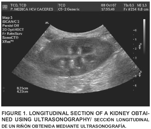
Transverse section. The animal was placed in lateral recumbency with the kidney to be examined uppermost. The transducer was positioned on the left flank just behind the last rib for the left kidney, and on the right flank, over the last two intercostal spaces, for the right kidney. Then, the kidney was located and the transducer was rotated through 90º to get the maximum transverse section. Renal width and height were measured in this plane (FIG. 1).
-
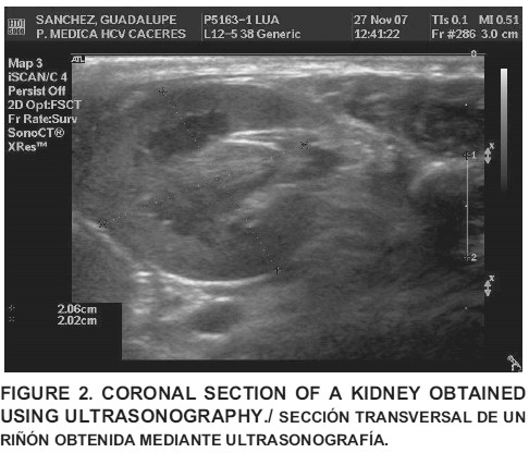
Coronal section. The animal was also positioned in lateral recumbency and the transducer situated in the location previously described. The transducer was moved until the long axis of the kidney was maximal. Renal length was recorded in this plane (FIG. 2).
Kidney volume determination using ultrasound
The approximate renal volume was estimated using the formula for the volume of an ellipsoid: V = L x W x H x 0.523 (V = volume; L = length; W = width; H = height).
Anatomic measurements
Renal length, width and height were measured using a caliper. Three measurements were performed for each parameter and the mean was recorded.
Anatomic volume determination
The anatomic volume was calculated using the means of the three direct measurements of renal length, width and height. The formula for the volume of an ellipsoid previously described (V = L x W x H x 0.523) was applied.
Statistical analysis
The repeatability of each method (ultrasonographic measurements and direct measurements) was calculated assessing the differences between pairs of repeated measurements and the mean of these differences (d) which is an estimate of the mean bias of the first experiment relative to the second. As a measure of repeatability, the British Standards Institution repeatability coefficient [8] was calculated as twice the SD of the differences. This coefficient is used as indication of the maximum difference likely to occur between repeated measurements.
A method agreement analysis between two protocols (ultrasonographic kidney dimensions and direct kidney dimensions) was done. Differences between paired measurements on the same samples and the mean of these differences (d) were calculated to estimate the mean bias of one method relative to the other. The 95% limits of agreement were calculated as d ± SD. SD is the standard deviation of the differences between paired measurements [8]. All calculations were done using Microsoft Excel [11].
RESULTS AND DISCUSSION
The physical examinations and laboratory work from the dogs used in this study were normal. The rest of results obtained are shown in TABLE I.
TABLE I. BODY WEIGHTS, ULTRASONOGRAPHIC AND ANATOMIC LINEAR DATAS AND RENAL VOLUME OF THE 19 DOGS. R = RIGHT; L = LEFT./PESO CORPORAL, MEDIDAS LINEALES ECOGRÁFICAS Y ANATÓMICAS Y VOLUMEN RENAL DE 19 PERROS. R = DERECHO; L = IZQUIERDO.
|
| Kidney parameters | ||||||||||||||||
|
|
| Ultrasonographic Measurements | Anatomic Measurements | ||||||||||||||
| No. Dogs | Weight (Kg) | Height (cm) | Width (cm) | Length (cm) | Volume (cm3) | Height | Width (cm) | Length (cm) | Volume (cm3) | ||||||||
|
|
| R | L | R | L | R | L | R | L | R | L | R | L | R | L | R | L |
| 1 | 5 | 2.5 | 2.4 | 2.4 | 2.3 | 5.3 | 4.2 | 17 | 12 | 2.8 | 2.4 | 2.2 | 2.1 | 3.9 | 4.2 | 13 | 14 |
| 2 | 25 | 4.4 | 3.5 | 4.1 | 3.4 | 6.9 | 7.1 | 65 | 43 | 3.5 | 3.5 | 3.6 | 3.4 | 5.1 | 6.6 | 33 | 44 |
| 3 | 8 | 3.5 | 3.5 | 3.7 | 2.9 | 5.4 | 5.5 | 37 | 29 | 5.4 | 3.5 | 3.5 | 2.8 | 5.5 | 5.6 | 54 | 27 |
| 4 | 26.8 | 4.2 | 4.9 | 4.1 | 4.2 | 7.7 | 7.9 | 70 | 86 | 4.2 | 4.9 | 3.2 | 3.7 | 7.7 | 7.4 | 55 | 57 |
| 5 | 16 | 4.6 | 3.5 | 3.6 | 3.3 | 7.5 | 5.6 | 66 | 34 | 4.5 | 3.5 | 3.5 | 3.6 | 7.5 | 7.4 | 61 | 66 |
| 6 | 37 | 4.2 | 4.1 | 3.9 | 3.4 | 8.1 | 6.6 | 70 | 48 | 3.9 | 4.1 | 3.3 | 3.2 | 7.2 | 8 | 49 | 46 |
| 7 | 26 | 3.5 | 4.3 | 5 | 3.6 | 6.4 | 6.6 | 58 | 54 | 5.9 | 4.3 | 4.3 | 3.8 | 8.7 | 8.5 | 114 | 87 |
| 8 | 29 | 4.6 | 4 | 3.9 | 4.7 | 7.7 | 8.3 | 73 | 81 | 3.9 | 4 | 3.4 | 3 | 8.1 | 8.3 | 54 | 64 |
| 9 | 18.9 | 4 | 3.7 | 3.6 | 3.2 | 6.2 | 6.1 | 46 | 39 | 3.5 | 3.7 | 2.6 | 2.5 | 6.6 | 6.1 | 31 | 28 |
| 10 | 29.7 | 4.7 | 5 | 3.8 | 5.1 | 8.7 | 8.8 | 81 | 117 | 5.1 | 5 | 3.9 | 4.4 | 9.3 | 9 | 97 | 107 |
| 11 | 41.25 | 4.2 | 4 | 3.7 | 3 | 8 | 6.3 | 65 | 39 | 4.6 | 4 | 3.4 | 3.3 | 8.3 | 8.4 | 67 | 75 |
| 12 | 41.9 | 4.4 | 4.3 | 4.3 | 3.5 | 8.6 | 8 | 85 | 63 | 5.3 | 4.3 | 3.4 | 2.8 | 8.4 | 7.1 | 77 | 56 |
| 13 | 28 | 3.3 | 4.7 | 3.2 | 3.8 | 6.9 | 7.4 | 39 | 69 | 3.9 | 4.7 | 3.6 | 3.5 | 8.1 | 8.1 | 60 | 66 |
| 14 | 27.3 | 4.3 | 4.5 | 3.5 | 3.7 | 7.2 | 7.5 | 57 | 64 | 5.3 | 4.5 | 3.6 | 2.8 | 8.4 | 7.1 | 82 | 56 |
| 15 | 17.7 | 3.3 | 3.5 | 2.8 | 2.7 | 5.7 | 6 | 28 | 30 | 3.2 | 3.5 | 2.4 | 2.5 | 5.7 | 5.6 | 23 | 22 |
| 16 | 47.5 | 4.8 | 5.5 | 4.2 | 4.9 | 7.5 | 9 | 78 | 126 | 5 | 5.5 | 4.1 | 4.6 | 10 | 10 | 109 | 135 |
| 17 | 7.4 | 2.7 | 2.9 | 2.2 | 2.3 | 5.5 | 4.5 | 17 | 15 | 2.8 | 2.9 | 2.2 | 2.3 | 5.1 | 4.9 | 17 | 18 |
| 18 | 7.1 | 2.9 | 4 | 2.4 | 2.6 | 5.6 | 5.2 | 20 | 9 | 2.6 | 4 | 2 | 2 | 5.3 | 4.9 | 14 | 13 |
| 19 | 29 | 4.7 | 2.5 | 5.1 | 3.9 | 8.1 | 8.1 | 101 | - | 4.8 | 2.5 | 3.8 | 4.2 | 8.7 | 8.8 | 83 | 95 |
The kidneys in the dog usually can be imaged from a ventral approach, with the animal situated in dorsal recumbency [1,2,3]. However, in this work both flanks have been used as acoustic windows to avoid the interference in renal measurements caused by gas bowel.
The aim of this study was to evaluate the accuracy and reproducibility of the linear measurements of canine kidneys using ultrasonography and to calculate the renal volume with the formula of an ellipsoid. Previous reports established the precision of both the ultrasonographic measurements of the kidney [1,2] and the estimation of the renal volume [3,6] for comparing with the direct measurements of the kidney. Because it has been shown that the kidney size changes following its removal from the body [6], in the present study, the difference with respect to previous report lies in the fact that the anatomic measurements from kidneys have been compared with the ultrasonographic measurements using the statistical method agreement (TABLE II). In addition, the renal volume was calculated from direct linear measurements obtained of the kidney (anatomic measurements), instead of using the classic method of water displacement; subsequently, the renal volume was compared with that obtained from the linear ultrasonographic measurements using as well the method agreement (TABLE II).
TABLE II. METHOD AGREEMENT ANALYSIS RESULTS./RESULTADOS DEL ANÁLISIS DE CONCORDANCIA ESTADÍSTICA.
| Kidney | Width (cm) | Height (cm) | Length (cm) | Volume (cm3) |
| Left | -0.32 ± 1.02 | 0.32 ± 1.86 | 0.39 ± 1.78 | 6.24 ± 54.60 |
| Right | -0.39 ± 0.88 | 0.16 ± 1.64 | 0.25 ± 2.16 | 1.15 ± 42.08 |
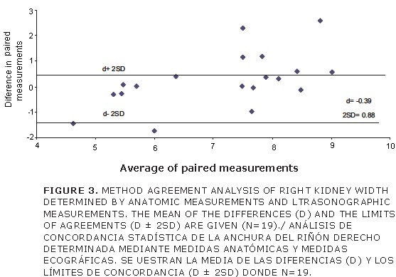
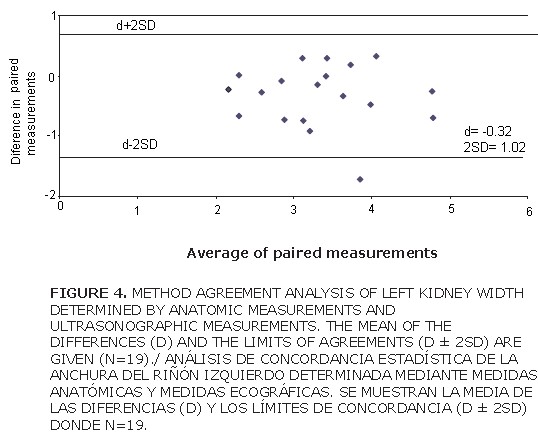
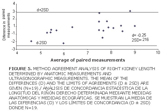
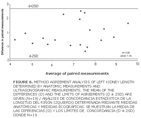
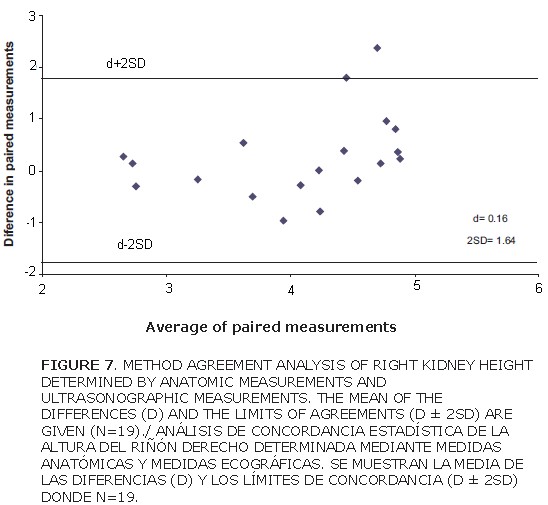
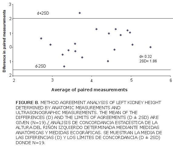
The difference between the means of the repeated measurements of the right kidney width was d = – 0.39 (TABLE II), and the value of the left kidney was d = – 0.32 (TABLE II). The British Standards Institution repeatability coefficients were 2SD = 0.88 (TABLE II) for the right kidney, and 2SD = 1.02 (TABLE II) for the left kidney (FIGS. 3 and 4) the length of each kidney measured in vivo was compared with the ultrasonographic dimensions. The mean difference between repeated measurements of the right kidney was d = 0.25 (TABLE II), with the British Standards Institution repeatability coefficient being 2SD = 2.16 (TABLE II and FIG. 5). The results for the left kidney were d = 0.39 and 2SD = 1.78 (TABLE II and FIG. 6). When the anatomic height measurement was compared with the ultrasonographic height measurements, the values of d were 0.16 for the right kidney (TABLE II) and 0.32 for the left kidney (TABLE II). The British Standards Institution repeatability coefficients were 2SD = 1.64 and 2SD = 1.86 for the right and left kidney, respectively (TABLE II and FIGS. 7 and 8).
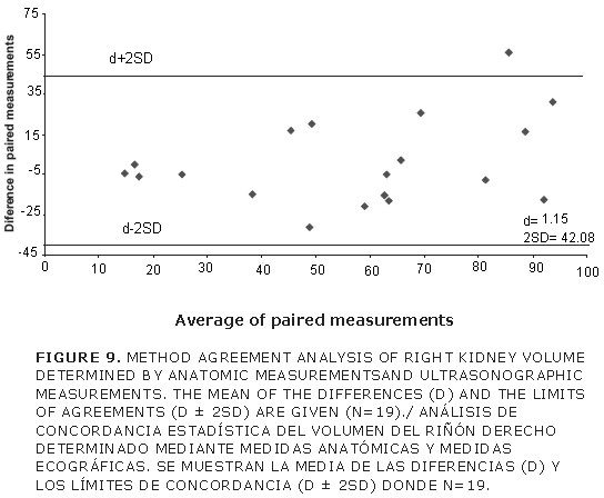
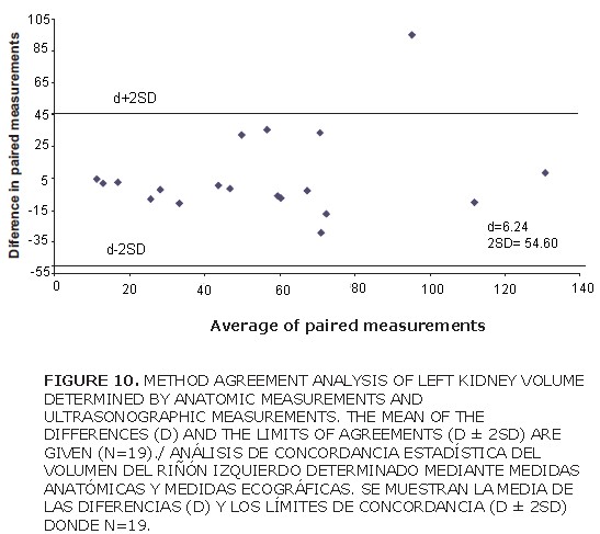
The limits of agreement (d ± 2 SD) of each volume kidney were calculated and the result for the right kidney was 1.15 ± 42.08 (TABLE II and FIG. 9), and 6.24 ± 54.60 for the left kidney (TABLE II and FIG. 10).
Two ways of evaluating renal measurement: ultrasonographic kidney dimensions and direct kidney dimensions were used. FIGS. 3 and 10 show that the scatter of the points is random, indicating that the size of the discrepancy between the two methods of measures is not related to the number of measurements. It was observed that 95% of differences lie within of limits of agreement, except for the width values from the right kidney. Right kidney ultrasound measurements are usually more difficult to obtain than those of the left kidney because of gas bowel and the cranial location of the right kidney under the rib cage.
CONCLUSION
This is a satisfactory agreement, and the ultrasonographic method is able to use for renal measures with reasonable confidence in the dogs. Therefore, it was concluded that linear kidney ultrasound measurements in vivo appear to be sufficiently accurate for clinical use. Renal volume may be calculated using the formula for the volume of an ellipsoid, and this gives a good indication of the true renal volume The exactitude of the measure depend of operators competence using ultrasound equipment and to the sonologist knowledge and experience.
BIBLIOGRAPHICS REFERENCES
1. BARR, F.J. Evaluation of ultrasound as a method of assessing renal size in the dog. J. Small Anim. Pract. 31: 174-179. 1990. [ Links ]
2. BARR, F.J.; HOLT, P.E.; GIBBS, C. Ultrasonograpic measurement of normal renal parameters. J. Small Anim. Pract. 31: 180-184. 1990. [ Links ]
3. FELKAI, C.S.; VOROS, K.; VRABELY, T.; KARSAL, F. Ultrasonographic determination of renal volume in dog. Vet. Radiol. Ultrasound. 33: 292-296. 1992. [ Links ]
4. KONDE, L.J. Sonography of the kidney. Vet. Clin. N. Am-Small. 15: 1149-1158. 1985. [ Links ]
5. LERMAN, L.O.; BENTLEY, M.D.; BELL, M.R.; BUMBERGER, J.A.; ROMERO, J.L. Quantitation of the in vivo kidney volume with cine computed tomography. Invest. Radiol. 25: 1206-1211. 1990. [ Links ]
6. NYLAND, T.G.; KANTRWITZ, B.M.; FISHER, P.E.; OLEANDER, H.J.; HORNOF, W.J. Ultrasonic determination of kidney volume in the dog. Vet. Radiol. 30:174-180. 1989. [ Links ]
7. NYLAND, T.G.; MATTON, J.S.; HERRGESELL, E.J.; WISNER, E.R. Urinary tract. In: Nyland, T.G., Matton, J.S. (Eds). Small Animal Diagnostic. Ultrasound. W B Sounders Company, Philadelphia, USA. 166-204 pp. 2002. [ Links ]
8. PETRIE, A.; WATSON, P. Diagnostic test. In: Statistics for Veterinary and Animal Science. Blackwell Science Ltd, London. 168-181pp. 1999. [ Links ]
9. SAMPAIO, K.M.O.R.; ARAUJO, R.B. Ultrasonographic measurement of linear characteristics and renal volumetry in dogs. Arq. Bras. Med. Vet. Zoo. 54: 50-54. 2002. [ Links ]
10. TERRIER, F.; HRICAK, H.; REVEL, D. Magnetic resonance imaging in the diagnosis of acute renal allograft rejection and its differentiation from acute tubular necrosis. Experimental study in the dogs. Invest. Radiol. 20: 617-625. 1985. [ Links ]
11. TRAVERÍA, S.; PRATS, C. Microsoft Excel 2003. Infor Books editions. 2004. [ Links ]
12. TWARDLOCK, A.R.; KRAWIEC, D.R.; LAMB, C.R. Kidney Scintigraphy. Seminars in Veterinary Medicine Surgery (Small Animal) W B Sounders Company, Philadelphia, USA. 6: 164-169. 1991. [ Links ]


 uBio
uBio 
